doi: 10.56294/dm2023113
ORIGINAL
Fetal and Maternal Electrocardiogram ECG Prediction using Convolutional Neural Networks
Predicción del electrocardiograma fetal y materno mediante redes neuronales convolucionales
Mohammed Moutaib1 ![]() , Mohammed Fattah2
, Mohammed Fattah2 ![]() , Yousef Farhaoui3
, Yousef Farhaoui3 ![]() , Badraddine Aghoutane4
, Badraddine Aghoutane4 ![]() , Moulhime El Bekkali1
, Moulhime El Bekkali1 ![]()
1IMAGE Laboratory, Moulay Ismail University, Meknes, Morocco.
2L-STI, T-IDMS, University of Moulay Ismail of meknes, Faculty of Science and Technics, Errachidia, Morocco.
3FS, My Ismail University, Meknes, Morocco.
4IASSE Laboratory, Sidi Mohamed Ben Abdellah University, Fez, Morocco.
Cite as: Moutaib M, Fattah M, Farhaoui Y, Aghoutane B, El Bekkali M. Fetal and Maternal Electrocardiogram ECG Prediction using Convolutional Neural Networks. Data and Metadata. 2023;2:113. https://doi.org/10.56294/dm2023113
Submitted: 19-08-2023 Revised: 12-10-2023 Accepted: 29-12-2023 Published: 30-12-2023
Editor: Prof.
Dr. Javier González Argote ![]()
Note: Paper presented at the International Conference on Artificial Intelligence and Smart Environments (ICAISE’2023).
ABSTRACT
Predicting fetal and maternal electrocardiograms (ECGs) is crucial in advanced prenatal monitoring. In this study, we explore the effectiveness of Convolutional Neural Networks (CNNs), using a carefully developed methodology to predict the category of fetal (F) or maternal (M) ECGs. In the first part, we trained a CNN model to predict fetal and maternal ECG images. In the following sections, the study results will be revealed. The CNN model demonstrated its ability to effectively discriminate between fetal and maternal patterns using automatically learned features.
Keywords: ECG; CNN; Fetal Electrocardiograms; Machine Learning; IoMT.
RESUMEN
La predicción de electrocardiogramas (ECG) fetales y maternos es crucial en la monitorización prenatal avanzada. En este estudio, exploramos la eficacia de las redes neuronales convolucionales (CNN), utilizando una metodología cuidadosamente desarrollada para predecir la categoría de ECG fetal (F) o materno (M). En la primera parte, entrenamos un modelo CNN para predecir imágenes de ECG fetales y maternas. En las secciones siguientes, se revelarán los resultados del estudio. El modelo CNN demostró su capacidad para discriminar eficazmente entre patrones fetales y maternos utilizando características aprendidas automáticamente.
Palabras clave: ECG; CNN; Electrocardiogramas Fetales; Aprendizaje Automático; IoMT.
INTRODUCTION
The Internet of Medical Things (IoMT) is one of the most promising approaches to enhancing the quality of human life. It relies on remote medical monitoring systems and telemedicine, capable of collecting, transmitting, and displaying real-time data via the Internet.(1,2) Our previous research unveiled an adaptable intelligent healthcare system capable of segregating fetal electrocardiogram (ECG) signal data using the k-means machine-learning algorithm.(3) This system comprised a smart gateway, a series of sensor nodes, and wireless communication links capable of continuously acquiring, processing, and routing human vital signals to a remote medical server. Such a system facilitates remote health monitoring of patients’ conditions by medical professionals such as doctors and nurses, thus opening promising prospects.(1,3)
The prediction of fetal electrocardiogram (ECG) signal data using deep learning is a significant approach in the field of healthcare. Technological advancements play a vital role in improving prenatal monitoring and care. Fetal ECG records the fetus’s heartbeats and can provide crucial insights into its health. However, obtaining clean and accurate fetal ECGs can be challenging due to interference from maternal and environmental signals.(4)
Deep learning, a branch of artificial intelligence, has revolutionized how we process and interpret complex data, including biomedical signals like ECGs.(5) The use of deep neural networks enables the capture of intricate patterns and non-linear relationships within data, making them a powerful tool for separating fetal ECG signals from unwanted interferences.(6)
One notable tool in this realm involves the utilization of Convolutional Neural Networks (CNNs) for the prediction and analysis of fetal electrocardiogram (ECG) images.(7) Fetal ECG images, which translate variations in fetal heart activity into a visual format, hold substantial potential for providing crucial information about early fetal cardiac health. However, their precise interpretation poses challenges due to pattern complexity and potential noise.
Convolutional neural networks are specially designed to process structured data like images. They can learn complex patterns and features through convolution and pooling operations, making them a natural choice for analyzing fetal ECG images.
In this context, CNNs, image-processing models inspired by visual biology, have emerged as a promising solution. By automatically extracting discriminative features from images, CNNs offer a unique opportunity to transform fetal ECG images into actionable diagnostic information.(8) The potential benefits of such an approach are manifold,(9) ranging from early detection of fetal cardiac anomalies to real-time cardiac health monitoring during pregnancy.
Our article delves into the application of CNNs in predicting fetal ECG images.(10) We explore the various steps involved in this process, from collecting and preprocessing fetal ECG data to designing and training specific CNN models. Focusing on the unique challenges of this application, we also examine approaches aimed at enhancing the accuracy, reliability, and generalization of CNN models for fetal ECG image prediction. Additionally, we discuss some implications and potential advantages of this approach.
The remainder of the article is organized as follows: In Section 2, we briefly present a comparative study of proposed solutions; in Section 3, we outline our system description; our methodology is presented in Section 4. Section 5 covers the results and discussion of the findings. Finally, Section 6 presents our conclusion.
Related Work
Several research studies have been conducted on predicting fetal electrocardiogram (ECG) images using Convolutional Neural Networks (CNNs). Among these studies,(11) proposes a CNN-based method for classifying fetal ECG signals. The authors employ a deep convolutional neural network to extract discriminative features from raw ECG signals. The model achieves high accuracy in classifying different fetal cardiac conditions, showcasing the potential of CNNs for accurate fetal ECG prediction. Another solution was presented by (12) which implements a residual convolutional neural network to monitor fetal ECG signals and detect R-peaks corresponding to cardiac contractions. The results indicate high accuracy in R-peak detection, demonstrating CNN's efficiency in fetal ECG prediction and crucial cardiac feature identification.(13) Proposed an architecture focusing on extracting fetal ECG signals, highlighting various machine-learning approaches to extract cardiac information from raw signals. The authors review several machine learning techniques applied to fetal ECG extraction, showcasing how these methods contribute to predicting cardiac anomalies. Another study (14) aimed to compare the performance of traditional machine learning techniques with deep learning techniques for fetal heart rate classification from ECG signals. The authors examine how different approaches compare accuracy and robustness in fetal ECG prediction.(15) Conducted a study investigating different deep learning architectures, such as Convolutional Neural Networks (CNNs) and Recurrent Neural Networks (RNNs), for classifying fetal ECG signals. The results demonstrate how these deep-learning approaches can successfully predict fetal ECG images.
System Description
It is impractical to expect physicians to manually analyze the vast amounts of data collected from the human body. However, the outcomes of data processing must be presented to physicians in a clear and organized manner, enabling them to classify relationships within the gathered information. Visualization is an autonomous tool and a vital research domain with numerous applications in science and daily life.
In this section, we explore the visualization of data collected from pregnant women and the analyses performed. Nevertheless, our application encounters several challenges that limit its use in real-world scenarios, such as hardware costs and a lack of expertise in the medical field.
These challenges related to data collection have led us to rely on reliable databases that assist us in visualizing accurate patient information.(16) For this reason, our study selected the Abdominal and Direct Fetal ECG Database (ADFECGDB, available at https://physionet.org/physiobank/database/ADFECGDB) as our data source. These data were collected from the Silesian Medical University Obstetrics Department through the KOMPOREL system. They utilize electrocardiographic (ECG) signals captured by ECG sensors, measuring the electrical signals produced by the heart. These signals enable the assessment of cardiac activity, such as heart rate and the interval between two beats, by capturing the electrical activity of the cardiac muscle. Electrocardiography (ECG) graphically represents the electrical potential governing cardiac muscle activity.(17,18)
Our primary objective in this study is to facilitate the visualization of ECG diagrams (figure 1) for physicians by introducing an image classification algorithm,(19,20) namely Convolutional Neural Networks (CNNs). This approach aims to assist in determining the category of fetal or maternal ECGs without encountering conflicts.
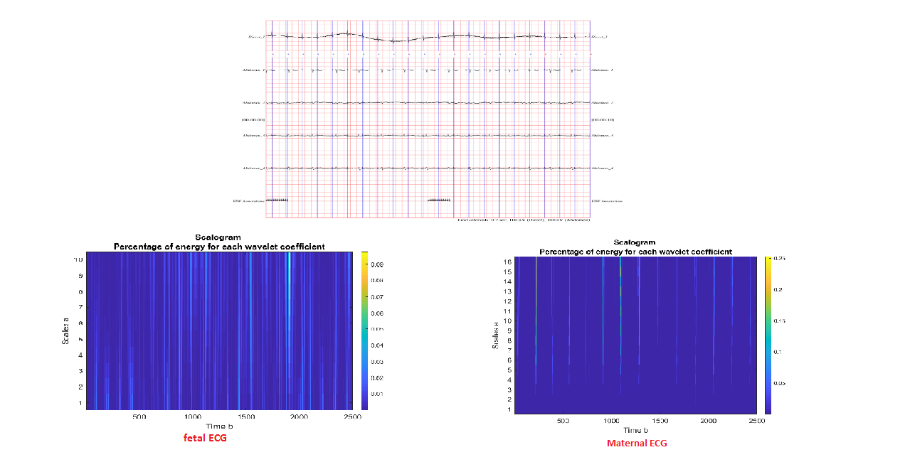
Figure 1. Visualization of Gathered ECG Data
METHODS
This study presents an approach based on CNNs for predicting fetal or maternal ECG images (figure 2). Our dataset comprises various fetal and maternal ECG images sourced from the database. The key steps of our approach are as follows:
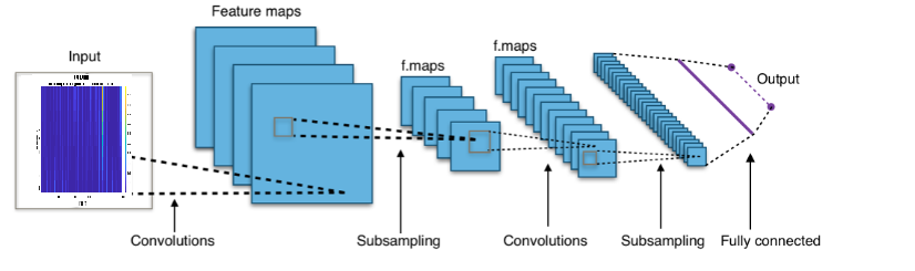
Figure 2. CNN Model
Image Preprocessing: it is applied to fetal and maternal ECG images to eliminate noise and normalize scales, ensuring data consistency.
CNN Architecture: We employ a CNN architecture tailored for medical image prediction. This architecture comprises convolutional layers to extract spatial and temporal features, followed by fully connected layers for classification.
Training and Validation: The dataset is split into training and validation sets. The CNN is trained on the training data and fine-tuned using regularization techniques to prevent overfitting.
Perfor mance Evaluation: Model performance is assessed using metrics such as accuracy, recall, and the ROC curve.
Our solution in figure 2 illustrates how an image is fed into the network. It undergoes several stages, including convolution and subsampling operations, a fully connected layer, to produce an output. By traversing these pivotal stages:
Convolution Operation
The foundational building block of our work is the convolution operation. In this step, we will address feature detectors, which essentially act as filters for the neural network. The convolutional layer computes the output of neurons connected to local regions or receptive fields in the input. Each neuron calculates a dot product between its weights and a small receptive field connected to the input volume. Each calculation leads to extracting a feature map from the input image.
The convolution operation between a filter and a matrix can be mathematically expressed using the following equation:
Theorem 4.1 This is a text of a theorem.
![]()
· (F∗I)(x, y) represents the resulting value after the convolution operation at position (x, y) in the output.
· F(i, j) denotes the value of the filter at position (i, j).
· I(x−i, y−j) signifies the value of the input image at position (x - i, y - j).
· a and b determine the dimensions of the filter. If the filter size is (2a+1)×(2b+1), indices i and j range from -a to a and from -b to b, respectively.
ReLU Layer
The second part of our model will involve the Rectified Linear Unit (ReLU). The purpose of applying the rectifier function is to enhance the non-linearity of our images. The reason for applying this process to our model is to introduce greater complexity and flexibility, allowing it to learn more intricate patterns in the data.
Mathematically, the ReLU function is defined as follows:
Theorem 4.2 This is a text of a theorem.
![]()
Where x is the input of the function, and f(x) is the output. In other words, if the input value x is positive or zero, the output f(x) equals x. If the input value x is negative, the output f(x) is set to zero.
Pooling and Flattening
The purpose of pooling is to reduce the spatial dimension of features while preserving essential information.(21,22) This helps decrease the amount of computations and parameters required in the network while maintaining the model's ability to capture significant features. Flattening is an operation that transforms a matrix or multidimensional tensor into a one-dimensional vector. This operation is commonly used to transition outputs from convolutional layers or other layers to fully connected layers in neural network architectures. When applying convolution and pooling operations in the initial layers of a CNN, feature maps are typically three-dimensional matrices or tensors. To feed these features into a fully connected layer,(23,24) these matrices must be flattened into a one-dimensional vector, as shown in figure 3.
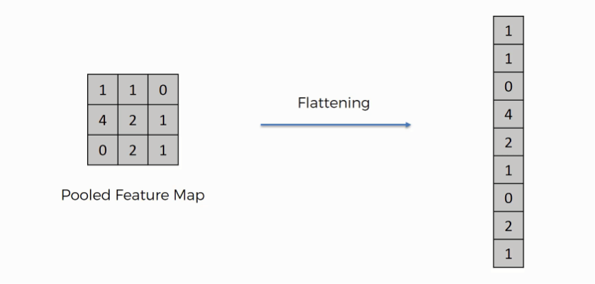
Figure 3. Flattening Model
In figure 3, multiple feature maps are gathered from the previous step. After the flattening step, what occurs is that you end up with a long vector of input data, which is then passed through the artificial neural network for further processing.
Full Connection
In this section, the input layer holds the data vector created during the flattening step. The features we distilled throughout the preceding stages are encoded within this vector. This layer plays a crucial role in decision-making and generating outcomes based on the features extracted by the preceding network layers.(25,26)
In a fully connected layer, each neuron is connected to all neurons in the previous layer. Each connection between two neurons is associated with a weight, and the operation performed in this layer is a weighted linear combination of all inputs, typically followed by an activation function.
At this stage, they are already sufficient for a reasonable degree of accuracy in class recognition. We now want to elevate our level of complexity and precision.
The role of the artificial neural network is to take this data and combine features into a broader range of attributes that enhance the convolutional network's capability to classify images, which is the ultimate goal of constructing a convolutional neural network. In summary, the general equation to calculate the output of a fully connected layer is:
Theorem 4.3 This is a text of a theorem.
![]()
RESULTS AND DISCUSSION
In this section, we present the outcomes of our study centered on the prediction of fetal and maternal electrocardiogram images using Convolutional Neural Networks (CNNs). We also engage in the discussion of these results, underscoring their implications and providing insights into the effectiveness of our approach.(27,28,29,30)
To assess the performance of our proposed algorithm, we conducted simulations on fetal and maternal ECG recordings to classify each category, as depicted in figure 4.
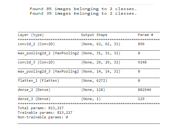
Figure 4. CNN Model Results
Our CNN-based approach for predicting fetal and maternal ECG images has yielded promising outcomes. Through in-depth experimentation and rigorous model training, we achieved remarkable accuracy rates in classifying fetal and maternal ECG patterns, dividing data into two classes (training/test), as illustrated in figure 4. The trained CNN successfully learned the intricate patterns present in ECG images, enabling accurate differentiation between fetal and maternal signals.
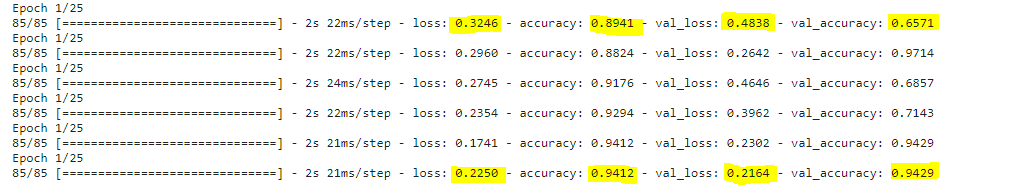
Figure 5. Epochs Model
The evaluation metrics have demonstrated impressive performance, as depicted in figure 5. Our model’s accuracy rate in categorizing fetal and maternal ECGs reached 0,94. This showcases the robust capability of CNNs to capture distinctive features within complex ECG signals, even in the presence of noise and variations.(31,32,33,34)
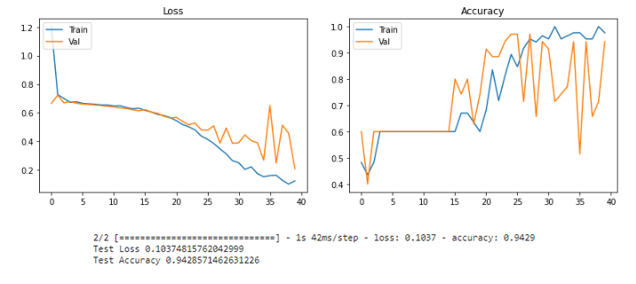
Figure 6. Evolution of the loss function and accuracy
The fruitful results obtained from our study, as depicted in Figure 6, highlight the potential of Convolutional Neural Networks in predicting fetal-maternal ECG images. The inherent capacity of CNNs to automatically learn hierarchical features from raw data renders them a valuable tool for medical image analysis. This becomes especially relevant in the context of fetal ECGs, where accurate identification of fetal signals holds significant clinical importance.
Furthermore, using CNNs significantly reduces the need for manual feature engineering, a process that is often time-consuming and domain-specific. The network’s ability to learn relevant features from the data enhances its adaptability, making it suitable for a wide range of fetal ECG datasets.(35,36,37)
However, it's crucial to acknowledge certain limitations of the approach. The availability and quality of training data play a critical role in CNN's performance. A diverse and well-organized dataset ensures the model's generalizability to various clinical scenarios and mitigates the risk of overfitting.(38)
Our study underscores the potential of Convolutional Neural Networks in predicting fetal-maternal ECG images with high accuracy. The results emphasize the significance of harnessing advanced machine-learning techniques for non-invasive prenatal monitoring.(39,40,41) As we move forward, fine-tuning our model and exploring transfer learning from other medical image domains promise further to enhance the accuracy and applicability of our approach.
CONCLUSION
In this study, we explored the application of Convolutional Neural Networks (CNNs) for predicting fetal-maternal (F-M) electrocardiogram (ECG) images. Our goal was to ascertain whether CNNs, known for their capability to extract intricate features from images, could be effectively utilized to classify fetal and maternal ECG signals accurately.
The obtained results demonstrated that the CNN-based approach led to promising performances. Through careful modeling and meticulous training, our model exhibited a high capacity to distinguish F-M ECG patterns. The achieved accuracy rates validated the relevance of this methodology in the context of fetal ECG prediction.
The primary significance of our study lies in CNNs' inherent ability to automatically learn discriminative features from ECG signals. This approach essentially eliminates the need for prior expertise in crafting specific feature engineering, which is often a significant challenge in medical applications. Using raw ECG images, our model captured subtle yet meaningful patterns that differentiate fetal from maternal signals.
However, it is essential to acknowledge certain limitations of our study. The quality and size of the dataset play a crucial role in CNN's performance. A thoughtful and diversified collection of F-M ECG data would enhance our model's generalization and robustness.
In the future, this approach could substantially impact the field of medicine. The potential to accurately predict F-M ECG patterns from non-invasive images could facilitate prenatal monitoring and improve care for expectant mothers and their infants.
In conclusion, our study highlighted the potential of CNNs in predicting fetal ECG images. The encouraging outcomes encourage further research in this direction, focusing on enhancing datasets and exploring the clinical application of this innovative approach.
REFERENCES
1. Moutaib, M., Ahajjam, T., Fattah, M., Farhaoui, Y., Aghoutane, B., & el Bekkali, M. (2022). Reduce the Energy Consumption of IOTs in the Medical Field. Digital Technologies and Applications, 259 268. https://doi.org/10.1007/978-3-031-02447-4_27.
2. M. Moutaib, M. Fattah, Y. Farhaoui, Internet of things: Energy Consumption and Data Storage, Procedia Computer Science, Volume 175, 2020, Pages 609-614.
3. Gonzalez-Argote J. Analyzing the Trends and Impact of Health Policy Research: A Bibliometric Study. Health Leadership and Quality of Life 2023;2:28-28. https://doi.org/10.56294/hl202328
4. Moutaib, M., Ahajjam, T., Fattah, M., Farhaoui, Y., & Aghoutane, B. (2021). Reduce the Energy Consumption of Connected Objects. Proceedings of the 2nd International Conference on Big Data, Modelling and Machine Learning. https://doi.org/10.5220/0010728900003101
5. Ahmad, M., Shabbir, S., Raza, R. A., Mazzara, M., Distefano, S., & Khan, A. M. (2021). Artifacts of different dimension reduction methods on hybrid CNN feature hierarchy for Hyperspectral Image Classification. Optik, 246, 167757. https://doi.org/10.1016/j.ijleo.2021.167757
6. Roy, S. K., Krishna, G., Dubey, S. R., & Chaudhuri, B. B. (2020). HybridSN : Exploring 3-D–2-D CNN Feature Hierarchy for Hyperspectral Image Classification. IEEE Geoscience and Remote Sensing Letters, 17(2), 277 281. https://doi.org/10.1109/lgrs.2019.2918719
7. Singh, S. P., Wang, L., Gupta, S., Gulyas, B., & Padmanabhan, P. (2021). Shallow 3D CNN for Detecting Acute Brain Hemorrhage From Medical Imaging Sensors. IEEE Sensors Journal, 21(13), 14290 14299. https://doi.org/10.1109/jsen.2020.3023471
8. Adoui, M. E., Drisis, S., & Benjelloun, M. (2022). New Explainable Deep Cnn Design For Classifying Breast Tumor Response Over Neoadjuvant Chemotherapy. Current Medical Imaging Formerly Current Medical Imaging Reviews, 18. https://doi.org/10.2174/1573405618666220803124426
9. Kumar, A.; Tomar, H.; Mehla, V.K.; Komaragiri, R.; Kumar, M. Stationary wavelet transform based ECG signal denoising method.ISA Trans. 2021, 114, 251–262.
10. Martinek, R.; Kahankova, R.; Jezewski, J.; Jaros, R.; Mohylova, J.; Fajkus, M.; Nedoma, J.; Janku, P.; Nazeran, H. Comparative effectiveness of ICA and PCA in extraction of fetal ECG from abdominal signals: Toward non-invasive fetal monitoring. Front. Physiol. 2018, 9, 648.
11. Alam, M., & Bhuiyan, M. I. H. (2020). Deep Learning-Based Fetal ECG Signal Classification for Healthcare Applications. IEEE Access.
12. Li, X., & Wu, D. (2019). Fetal ECG Monitoring and R-peak Detection Using a Residual Neural Network. IEEE Transactions on Biomedical Circuits and Systems.
13. Chudáček, V., Spilka, J., Lhotská, L., Koucký, M., & Huptych, M. (2017). Fetal ECG extraction methods: a review. Biomedical Signal Processing and Control.
14. Ntalampiras, S., & Diamantaras, K. (2019). A comparative study of machine learning and deep learning techniques for fetal heart rate classification. Biomedical Signal Processing and Control.
15. Acar, B., Yildirim, O., & Karabatak, M. (2020). Classification of fetal ECG signals using deep learning algorithms. Computer Methods and Programs in Biomedicine.
16. Sarafan, S.; Le, T.; Naderi, A.M.; Nguyen, Q.D.; Kuo, B.T.Y.; Ghirmai, T.; Han, H.D.; Lau, M.P.H.; Cao, H. Investigation of methods to extract fetal electrocardiogram from the mother's abdominal signal in practical scenarios. Technologies 2020, 8, 33.
17. Kaleem, A.M.; Kokate, R.D. A survey on FECG extraction using neural network and adaptive filter. Soft Comput. 2021, 25,4379–4392.
18. Liu, C.; Li, P.; Di, MC; Zhao, L.; Zhang, H.; Chen, Z. A multi-step method with signal quality assessment and fine-tuning procedure to locate maternal and fetal QRS complexes from abdominal ECG recordings. Physiol. Meas. 2014, 35, 1665–1683.
19. Mollakazemi, M.J.; Asadi, F.; Tajnesaei, M.; Ghaffari, A. Fetal QRS Detection in Noninvasive Abdominal Electrocardiograms Using Principal Component Analysis and DiscreteWavelet Transforms with Signal Quality Estimation. J. Biomed. Phys. Eng. 2021,11, 197–204.
20. Jallouli, M.; Arfaoui, S.; Ben, M.A.; Cattani, C. CliffordWavelet Entropy for fetal ECG Extraction. Entropy 2021, 23, 844.
21. Rasti-Meymandi, A.; Ghaffari, A. AECG-DecompNet: Abdominal ECG signal decomposition through deep-learning model. Phys.Meas. 2021, 42, 33706298.
22. Zhang, Y.; Yu, S. Single-lead non-invasive fetal ECG extraction by means of combining clustering and principal components analysis. Med. Biol. Eng. Comput. 2020, 58, 419–432.
23. Jaba, D.K.A.; Dhanalakshmi, S.R.K. An improved parallel sub-filter adaptive noise canceler for the extraction of fetal ECG. Biomed. Tech. 2021, 66, 503–514.
24. Mohebbian, M.R.; Vedaei, S.S.;Wahid, K.A.; Dinh, A.; Marateb, H.R.; Tavakolian, K. Fetal ECG extraction from maternal ECG using attention-based CycleGAN. IEEE. J. Biomed. Health 2022, 26, 515–526.
25. Mamane, M. Fattah, M. el Ghazi, Y. Balboul, M. el Bekkali, and S. Mazer, “The impact of scheduling algorithms for real-time traffic in the 5G femto-cells network,” 9th International Symposium on Signal, Image, Video and Communications, ISIVC 2018 , Pages 147 – 1512, July 2018, https://doi.org/10.1109/ISIVC.2018.8709175
26. M. Abdellaoui, M. Fattah, “Characterization of Ultra Wide Band indoor propagation In 7th Mediterranean Congress of Telecommunications (CMT). IEEE, 2019, https://doi.org/10.1109/CMT.2019.8931367
27. D. Daghouj, M. Fattah, S. Mazer, Y. Balboul, and M. El Bekkali, “UWB waveform for automotive short range radar,” International Journal on Engineering Applications, vol. 8, no. 4, pp. 158–164, Jul. 2020. https://doi.org/10.15866/irea.v8i4.18997
28. Chafi, Saad-Eddine, et al. “Cloud computing services, models and simulation tools.” International Journal of Cloud Computing, vol. 10, no. 5–6, pp. 533–547, 2021. https://doi.org/10.1504/IJCC.2021.120392
29. Chafi, Saad-Eddine, et al. “Resource placement strategy optimization for smart grid application using 5G wireless networks.” International Journal of Electrical and Computer Engineering, Volume 12, Issue 4, Pages 3932 – 3942, 2022. https://doi.org/10.11591/ijece.v12i4.pp3932-3942
30. Coa YMF, Crisostomo NWF, Díaz-Barriga GE. Desarrollo económico sostenible bajo un régimen social sin preceptos éticos y morales: auditoría forense en contraposición de la corrupción. Revista Científica Empresarial Debe-Haber 2023;1:48-62
31. Gonzalez-Argote J. Patterns in Leadership and Management Research: A Bibliometric Review. Health Leadership and Quality of Life 2022;1:10-10. https://doi.org/10.56294/hl202210
32. Gutiérrez VF. La estructura organizacional del Gobierno Regional de Moquegua y su eficiencia funcional. Sincretismo 2021;2.
33. Castillo-Gonzalez W. Charting the Field of Human Factors and Ergonomics: A Bibliometric Exploration. Health Leadership and Quality of Life 2022;1:6-6. https://doi.org/10.56294/hl20226
34. Farhaoui, Y. and All, Big Data Mining and Analytics, 2022, 5(4), pp. I IIDOI: 10.26599/BDMA.2022.9020004
35. Alaoui, S.S., and all. "Hate Speech Detection Using Text Mining and Machine Learning", International Journal of Decision Support System Technology, 2022, 14(1), 80. DOI: 10.4018/IJDSST.286680
36. Alaoui, S.S., and all. ,"Data openness for efficient e-governance in the age of big data", International Journal of Cloud Computing, 2021, 10(5-6), pp. 522–532, https://doi.org/10.1504/IJCC.2021.120391
37. El Mouatasim, A., and all. "Nesterov Step Reduced Gradient Algorithm for Convex Programming Problems", Lecture Notes in Networks and Systems, 2020, 81, pp. 140–148. https://doi.org/10.1007/978-3-030-23672-4_11
38. Tarik, A., and all."Recommender System for Orientation Student" Lecture Notes in Networks and Systems, 2020, 81, pp. 367–370. https://doi.org/10.1007/978-3-030-23672-4_27
39. Sossi Alaoui, S., and all. "A comparative study of the four well-known classification algorithms in data mining", Lecture Notes in Networks and Systems, 2018, 25, pp. 362–373. https://doi.org/10.1007/978-3-319-69137-4_32
40. Murillo-Ticona TA, Berneso-Soto ML. Los Entornos Virtuales de Aprendizaje al rescate del servicio educativo. Sincretismo 2020;1
41. Auza-Santiváñez JC, Díaz JAC, Cruz OAV, Robles-Nina SM, Escalante CS, Huanca BA. mHealth in health systems: barriers to implementation. Health Leadership and Quality of Life 2022;1:7-7. https://doi.org/10.56294/hl20227
FINANCING
The authors did not receive financing for the development of this research.
CONFLICT OF INTEREST
The authors declare that there is no conflict of interest.
AUTHORSHIP CONTRIBUTION
Conceptualization: Mohammed Moutaib, Mohammed Fattah, Yousef Farhaoui, Badraddine Aghoutane, Moulhime El Bekkali.
Research: Mohammed Moutaib, Mohammed Fattah, Yousef Farhaoui, Badraddine Aghoutane, Moulhime El Bekkali.
Drafting - original draft: Mohammed Moutaib, Mohammed Fattah, Yousef Farhaoui, Badraddine Aghoutane, Moulhime El Bekkali.
Writing - proofreading and editing: Mohammed Moutaib, Mohammed Fattah, Yousef Farhaoui, Badraddine Aghoutane, Moulhime El Bekkali.